X-rays and Scans for Dogs and Cats: How to Prepare
Verified by: Dr Luisa MRCVS
Published on: 25 September 2026
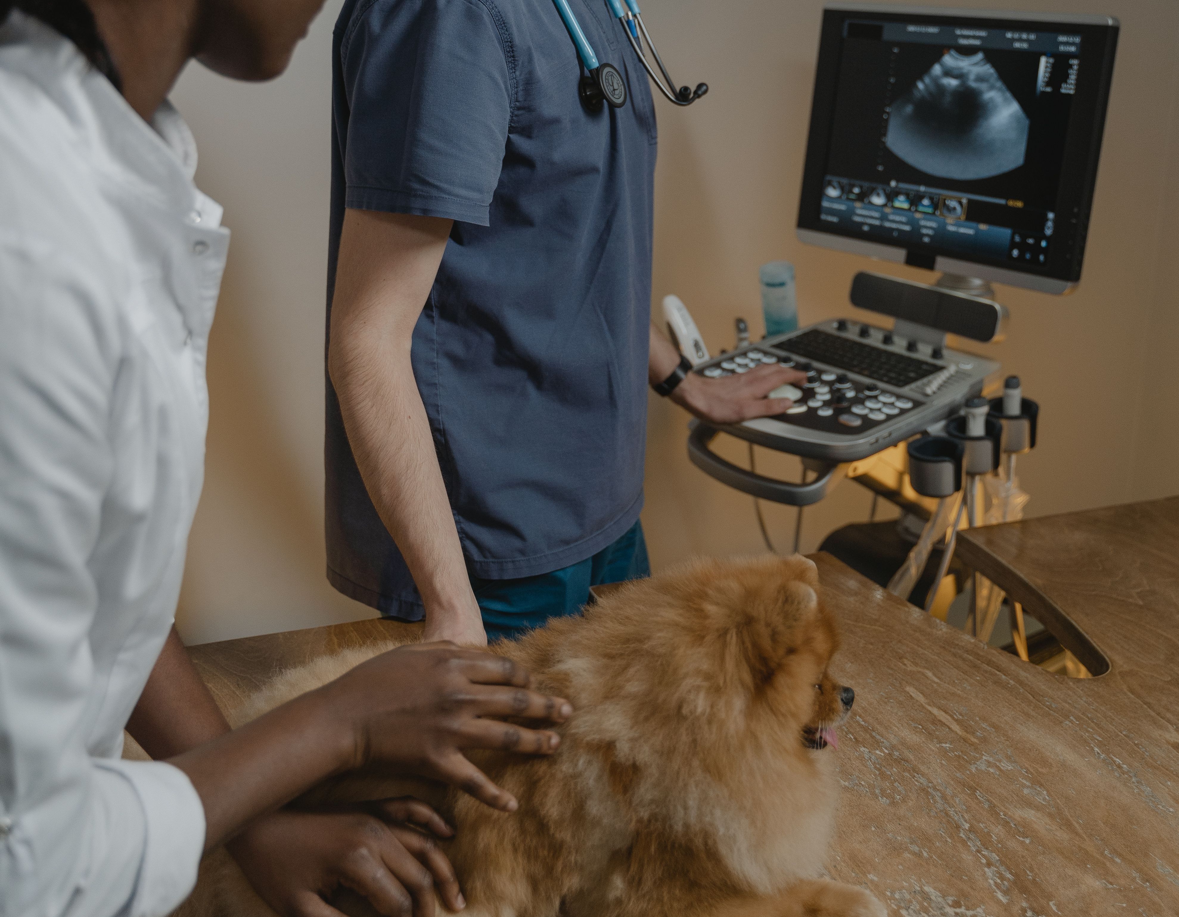
If your vet has recommended an X-ray or scan for your pet, it's natural to have a few questions. What are they looking for? Does your pet need to skip breakfast? What happens afterwards? This guide explains why imaging is used, how to get your dog or cat ready at home and what to expect next.
Why has my vet recommended imaging?
Diagnostic imaging lets your vet see inside your pet's body without surgery. It gives them a view of internal organs, bones and tissues so they can investigate symptoms and diagnose disease or other medical conditions.
Some problems simply can't be picked up during a physical examination. Imaging helps your vet spot structural changes that would otherwise go unnoticed, which means they can reach an accurate diagnosis and put together the most effective treatment plan for your pet.
How to prepare your dog for imaging
- Withhold food if your vet asks you to. You may be told not to feed your dog for 6 to 12 hours before the appointment. A full stomach can make it harder to get clear abdominal ultrasound images and can increase the risks if sedation or anaesthesia is needed.
- Keep water available. Make sure your dog has access to fresh water unless your vet has specifically told you otherwise.
- Go for a short walk before you arrive. This gives your dog the chance to wee and poo before their appointment. The exception is if your vet has also asked for a urine sample, in which case you'll want to save it! Our guide on how to collect a urine sample from your dog or cat explains how.
How to prepare your cat for imaging
- Withhold food if your vet asks you to. As with dogs, you may be told not to feed your cat for 6 to 12 hours beforehand. A full stomach can affect abdominal ultrasound images and increase the risks if sedation or anaesthesia is needed.
- Keep your cat indoors the night before. This stops them wandering off or finding something to eat outside, which could undo all your fasting efforts.
- Make the journey as calm as possible. Use a secure, comfortable carrier lined with familiar bedding to help keep stress to a minimum.
What types of imaging might my vet use?
There are several types of imaging and each is suited to looking at different parts of the body.
| Type | How it works | Best for |
|---|---|---|
| X-Ray | Low-dose radiation | Bones, joints, chest and lungs, foreign objexts in the gut |
| Ultrasound | High-frquency sound waves | Soft tissues, abdominal organs, the heart |
| CT scan | Advanced X-ray technology | Complex bony, nasal or internal structures |
| MRI | Magnetic fields and radio waves | Brain, spine and nervous system |